M-Eye Examinations and Testing

Header Logo

602-547-3255
patientcare@accenteyes.com

16020 North 35th Avenue
Suite 2
Phoenix, AZ 85053

OUR OFFICE HOURS

MON - THUR - 9:00AM - 6:00PM
TUE - 9:00AM - 6:30PM
FRI - 8:00AM - 2:30PM

  • Btn

  • Accent Eye Care M-Eye Examinations and Testing
  • Accent Eye Care M-Eye Examinations and Testing
  • Accent Eye Care M-Eye Examinations and Testing
  • Accent Eye Care M-Eye Examinations and Testing
  • Accent Eye Care M-Eye Examinations and Testing
Accent Eye Care M-Eye Examinations and Testing

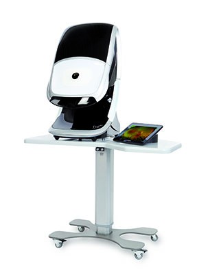
Pioneering Technology by Optos – Daytona Plus

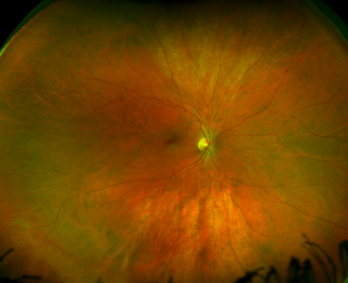
Optos’ patented ultra-widefield digital scanning laser technology acquires images of the back of the eye that support the detection, diagnosis, analysis, documentation and management of ocular pathology and systemic disease that may first present in the periphery. These conditions may otherwise go undetected using traditional examination techniques and equipment. Simultaneous, non-contact, central pole-to-periphery views of up to 82% or 200 degrees of the retina are displayed in one single capture, compared to 45 degrees achieved with conventional methods.

Accent Eye Care M-Eye Examinations and Testing

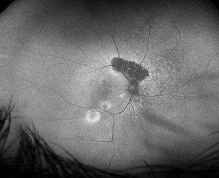
Play the video below to find out about the new technology of Autofluorescence in the Daytona Plus. This has the potential to discover pathology earlier than other standard retinal cameras because it can detect stressed retinal cells.

Accent Eye Care M-Eye Examinations and Testing

Glaucoma Screening Test GST from Haag Streit

Less than one minute 

Our new Glaucoma Screening Test GST can distinguish between normal and abnormal visual fields in less than one minute. The test is purely qualitative and distinguishes between normal and abnormal visual fields by presenting stimuli three times at a brightness that patients with normal vision should see. If not seen, then the visual field is flagged as abnormal with high reliability and the patient can be further tested. Because the test is efficient, it opens doors for more routine visual field testing to ensure no pathology goes undetected. The patient-friendly Pulsar stimulus has been developed for early glaucoma detection and shows a short learning curve and low test-retest variability.

Sensitive glaucoma analysis

The sensitive Cluster Analysis groups visual field defects along nerve fibre bundles and combines high sensitivity with good specificity to detect early glaucomatous changes. Significant defects are highlighted and the cluster defects can be used for structural comparison.

 For intuitive structural comparison

Combining the results of both structure and function is key to obtaining a comprehensive assessment of the onset and progression of glaucoma. The Octopus Polar Analysis projects local visual field defects along the nerve fibers to the optic disk and displays them oriented as structural results.

Accent Eye Care M-Eye Examinations and Testing

New Pediatric Technology – Plusoptix

Accent Eye Care now has the recently developed Plusoptix Automated Refractor used to detect most common vision disorders in children as early as possible.

In combination with other entry examination tests, Dr. Gong and her staff will be able to determine a valuable starting point for myopia, astigmatism and axis measurements (information used for prescribing eyewear).

Plusoptix pediatric auto-refractors take binocular (both eyes simultaneously) or monocular readings from 1 meter (3.3 feet) away in less than one second and can even be used for strabismic (eye turn) or aphakic (lens removed from eye) patients.

SD-OCT (Spectral Domain – Optical Coherence Tomography)

Accent Eye Care M-Eye Examinations and Testing

OCT provides cross-sectional images of the retina through retinal imaging. We have the newest spectral domain (SD) technology which improves image quality and acquisition, providing a larger set of data points for accurate spatial correlations and mapping of individual retinal layers.

Accent Eye Care M-Eye Examinations and Testing

This new technology allows us to easily conduct an eye exam in Phoenix, AZ by offering complete retina, glaucoma, and keratoconus/anterior segment scanning as part of our advanced diagnostic testing. Because the streamlined user interfaces are fast and efficient, you will not be waiting long for your results.

Topography

Accent Eye Care M-Eye Examinations and Testing

Corneal topography is a non-invasive medical imaging technique for mapping thousands of points of the corneal surface, the front of the eye. This can be crucial in determining the quality of vision for conditions such as keratoconus and pellucid marginal degeneration, which may exhibit corneal steepening before any biomicroscopic signs are evident. Topography also is invaluable when evaluating pre- and postsurgical patients, especially corneal transplants, radial keratotomy or LASIK. Postoperatively, topography can help follow the healing phase and assist with contact lens fitting, especially for hard to fit contacts.

Accent Eye Care M-Eye Examinations and Testing

New Technology for digital preliminary testing

With the newly developed Stereo Optical digital Optec Plus, Dr Gong’s office can efficiently test for: loss of focusing flexibility, clarity of vision at distance and near, astigmatism ( 2 prescriptions required, 90 degrees apart ), latent hyperopia (farsightedness), fusion (ability to use your eyes together), lateral and vertical phoria ( where the eyes rest horizontally and vertically), depth perception ( 3-D vision), Color vision tests, contrast sensitivity test ( your ability to tell the difference between the target and background) and Horizontal peripheral vision testing for the DMV paperwork: Stimuli at 45° nasal and 55°, 60°, 70°, 80°, 85°, 90° and 100° temporal.

Retinal Imaging

Digital retinal imaging is another non-invasive procedure used by our office to conduct that incorporates high-resolution imaging systems to take pictures of the nerve-rich lining of your eye that you use to see with, the retina. This helps us to detect and manage eye and health conditions such as glaucoma, diabetes, and macular degeneration.

Retinal images provide a permanent and historical record of changes in your eye. Images can be compared side-by-side, year after year, to discover even subtle changes and help monitor your eye health.

Accent Eye Care M-Eye Examinations and Testing

Gene testing

Gene testing is a procedure to identify early or intermediate age-related macular degeneration risks. Gene testing recognizes those who will most likely develop advanced macular degeneration with vision loss.

For the test, a simple cheek swab is taken in Dr. Gong’s office, and is at no cost to you if you have medical insurance. The only charge would be for the office visit to review the results.

Macular degeneration usually goes unnoticed in most people until they lose central vision in one eye. The loss of vision can happen rapidly and without warning; it may be detained but is typically irreversible. In order to prevent blindness from macular degeneration, early discovery and treatment is crucial.

Visual Field Analyzer

A visual field test in our AZ eye exam facility is designed to detect dysfunctions in central and peripheral vision which can be caused from various medical conditions such as glaucoma, stroke, brain tumors, and other neurological deficits.

Our eye exam in Phoenix, AZ is performed by using the Visual Field Analyzer from Carl Zeiss, which utilizes Frequency Doubling Technology (FDT). FDT is proven technology used to target low redundancy ganglion cells; because these cells are sparsely populated, there is less compensation available to mask cell damage. By targeting these cells, the Humphrey FDT perimeters can detect early visual field loss.

Accent Eye Care M-Eye Examinations and Testing

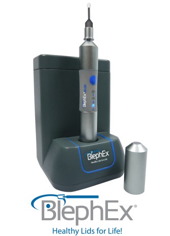
BlephEx – Blepharitis treatment

The reason that the BlephEx treatment is so successful, is that it completely removes the exotoxin/lipase/superantigen-laden biofilm along the lid margin. This biofilm has been building up on our patients lids for decades, causing low-grade inflammation and slowly turning down and degrading tear production.

For some of you who do Bleph Ex with us, when you leave our office, you will have completely clean lids, biofilm free for the first time in decades. This is the critical first step in effectively treating all forms of blepharitis and dry eye.

The biofilm is a necessary component for the Staph to become virulent. It is a multi-layered polysaccharide matrix with lots of channels, which allow for increased surface area as well as cell-to-cell communication. The density of bacteria can be greatly increased within this biofilm, which leads to quorum-sensing gene activation of virulence factors such as exotoxins, lipases and super-antigens.

Think of a low bacterial density, non-virulent, as a community of single family homes. The biofilm acts as a 20 story high-rise apartment complex, leading to a high density of bacteria, and thus virulence.

The BlephEx treatment is the “bulldozer and wrecking ball” that will destroy these apartment complexes, thus greatly decreasing the density, and therefore virulence of the bacteria.

So:

Biofilm=apartment complex

BlephEx=bulldozer